Intraoperative Visualization Improves Outcomes for Complex Spine Surgery
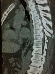
Duke spine surgeons are using mobile intraoperative computed tomography (CT) scanners to perform an increasing number of highly complex spine surgeries. The scanners offer many advantages over preoperative CT scans and intraoperative fluoroscopy for image guidance in complex cases.
“In patients with deformity, failed previous surgery, an injury, or a tumor, it’s often difficult to identify distorted anatomy,” says Duke spine surgeon Norah Foster, MD. The scanners allow surgeons to obtain 3-dimensional images of a larger field than other intraoperative modalities—including the entire spine from cranium to pelvis—to help ensure accurate placement of hardware.
Images can be obtained at any point before, during, or directly after surgery. By imaging the area immediately after the procedure, surgeons can confirm that the hardware placement is correct. “If a screw isn’t in an ideal position, we can reposition it immediately and avoid another trip to the operating room for the patient,” says Foster.
The scanners also support minimally invasive procedures by allowing surgeons to visualize the surgical field in 3 dimensions and accurately place instruments and hardware through small incisions with a high degree of accuracy.
Duke surgeons often use a sophisticated navigation system in combination with the scanners, placing sensors in a stable, static location (such as a bony structure) and using a probe with a sensor to track the location of surgical instruments in relation to the patient’s anatomy, helping them avoid critical structures.
Foster notes that there’s another advantage to using the intraoperative CT scanners instead of standard fluoroscopy and CT scans: less radiation exposure for surgical teams and possibly patients. “We often need to take multiple images using fluoroscopy during a procedure, and throughout a surgeon’s career, that can mean a lot of cumulative radiation exposure that may contribute to cataracts and certain cancers,” she says.
Although the scanners deliver approximately the same amount of radiation to patients as other CT scanners, Duke surgeons are finding that they can adjust the amount of radiation to as little as 25% of the normal dose from a CT scan and still obtain suitable images.
“This technology is the very definition of ‘novel,’ and it isn’t readily available at most hospitals. It’s a great example of how Duke is committed to having the most advanced resources possible to help patients with complex needs,” says Foster.