Powerful Imaging Technologies Offer Early Stage Diagnoses
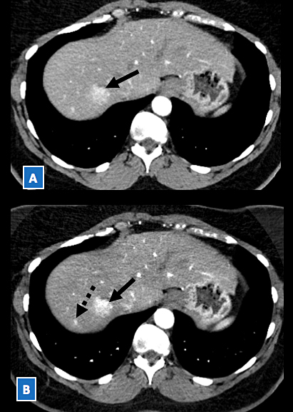
FIGURE 1. DECT images in a patient with metastatic disease to the liver. A) Image obtained during late hepatic arterial phase with a conventional CT protocol reveals a faint 2.5-cm hypervascular lesion (arrow) in the right liver lobe, which is barely visible against the background liver. B) Corresponding image obtained using a dual-energy acquisition demonstrates markedly increased conspicuity (ie, visibility) of this lesion (arrow) against the background liver, as well as a second smaller lesion (dashed arrow) in the posterior right liver lobe.
The use of dual-energy computed tomography (DECT) is growing as an efficient diagnostic tool, providing detailed information about early stage disease conditions.
“The DECT scan is a powerful tool that allows us to assess material composition to offer very specific diagnostic information,” says Daniele Marin, MD, a Duke abdominal imaging specialist. “We can better determine the aggressiveness of a tumor and provide a precise grade classification.”
The Duke Abdominal Imaging Division has been a leader in the clinical validation of DECT in the United States and is one of the few radiology centers in the Carolinas using DECT as a daily clinical tool.
The technology has specific applications in detecting and classifying gout crystals and kidney stones, Marin says. But those applications are just the tip of the iceberg: DECT’s most effective use, he says, includes early detection of diseases and improved characterization of incidental lesions. Routine use of DECT may eliminate the need for additional imaging in the workup of some of these incidental lesions (Figure 1).
DECT’s diagnostic value is evident when patients present with common but difficult-to-diagnose conditions such as abdominal pain. Computed tomography (CT) scans—the older, more common imaging technology—may identify a lesion but, unlike DECT, cannot provide detailed diagnostic information; multiple CT studies may be required to reach a definitive finding.
“The dual energy technology reduces the need for second CT scan studies,” Marin says. “By using 2 types of energy during the scan, the imaging is more precise, and the resulting studies are easier to interpret.” In addition, Marin says, the technology reduces radiation exposure to patients (Figure 2).
Although the imaging studies offer significant diagnostic advantages, the future growth of DECT is limited by the workflow challenges presented by the increase in images and data—a concern Duke is addressing as it looks toward expanding the use of the technology in the future.
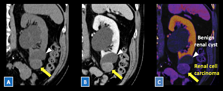
FIGURE 2. A, B) DECT images in a patient with renal cell carcinoma. C) The tumor (yellow arrow) demonstrates increased iodine content (ie, vascularity) on the color coded iodine overlay image. Also note the lack of iodine (ie, vascularity) within an adjacent benign simple renal cyst (white arrow).